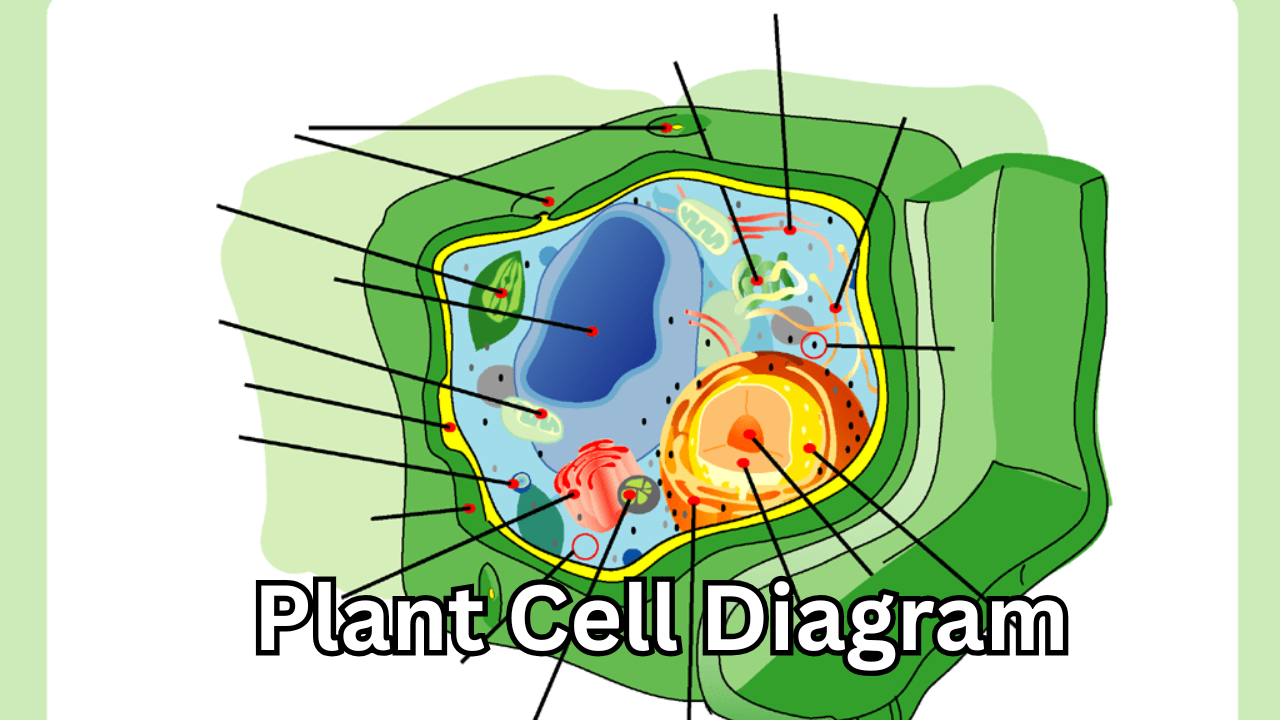
A plant cell diagram offers a clear and engaging way to visualize the structure and function of the basic unit of plant life, making it easier for students, teachers, and science enthusiasts to grasp complex biological concepts at a glance. By showing the placement of organelles and their relationships inside the cell, this visual representation turns abstract textbook descriptions into something concrete and meaningful, which is why it remains such an essential tool in biology education.
The importance of visualizing plant cells in biology
Understanding plant biology starts with understanding the cell. Every plant, whether a tiny blade of grass or a massive oak tree, is built from countless microscopic units that work together in harmony. These units are packed with specialized parts that perform different roles. Some produce energy, others store nutrients, or protect the cell’s interior. Without a visual reference, it can be difficult to imagine how all these components fit together inside such a tiny space.
That is where a detailed visual representation becomes valuable. By seeing how each organelle is positioned and how it interacts with others, learners gain a more complete picture of how plant life operates. Instead of memorizing lists of cell parts, they can understand the system as a whole. This leads to better retention and deeper comprehension. This visual approach also helps bridge the gap between theory and real-life biological processes, making science feel more intuitive and less intimidating.
How a plant cell is organized
Plant cells are remarkable for their level of organization. They are surrounded by a sturdy outer layer that provides protection and shape. This allows plants to stand upright and grow in defined forms. Inside this outer boundary is a flexible membrane that controls what enters and leaves the cell. The membrane ensures essential substances move in while waste products are removed.
Within this enclosed space lies a busy environment filled with specialized structures. Each one has a unique function that contributes to the plant’s survival and growth. Some parts focus on producing energy, others on creating food, and some on storing materials for later use. When all these elements work together, the cell can carry out the processes that sustain the entire plant.
Visual models clarify this organization. Seeing each component’s location makes it easier to understand the cell as a system rather than just a collection of parts.
The role of the nucleus in plant life
Most plant cells have a nucleus at the center, serving as the control hub. It contains genetic material that guides growth, development, and environmental responses, shaping leaves, flowers, and responses to sunlight.
The nucleus also directs the production of proteins, which are essential for nearly every cellular activity. These proteins help build structures, speed up chemical reactions, and carry out tasks that keep the cell functioning smoothly. By controlling these processes, the nucleus ensures the cell operates in accordance with the plant’s overall needs.
A visual layout that highlights the nucleus helps learners see why it is often called the “brain” of the cell. Its central location and connections to other structures make its importance clear.
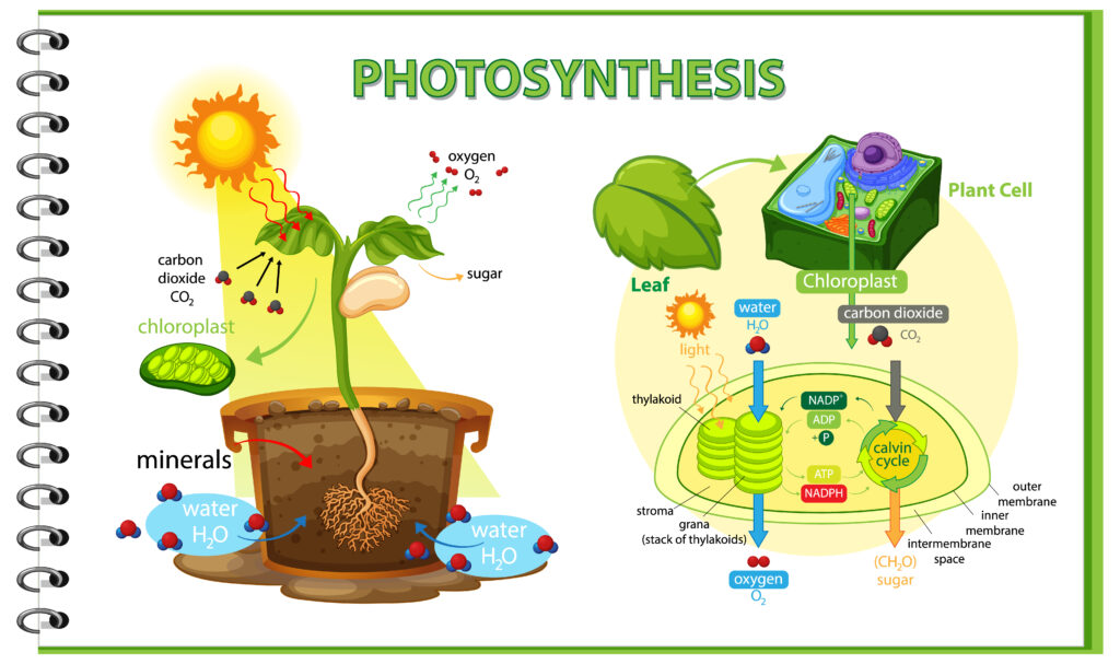
Chloroplasts and the magic of photosynthesis
One of the most distinctive features of plant cells is the presence of chloroplasts. These green-colored structures are responsible for photosynthesis. This is the process by which plants convert sunlight into usable energy. Using light, water, and carbon dioxide, chloroplasts produce glucose, which fuels the plant. They also release oxygen into the atmosphere.
This process is not only vital for plants but also for life on Earth. The oxygen we breathe and the food chains that sustain animals depend on photosynthesis occurring within these tiny organelles. Understanding where chloroplasts are located and how they function gives insight into how plants support entire ecosystems.
Seeing chloroplasts within the cell helps explain energy flow. It turns unseen chemistry into something real and understandable.
The central vacuole and cellular balance
Another important structure in plant cells is the large central vacuole. This compartment takes up a significant portion of the cell’s volume. It serves several key purposes. It stores water, nutrients, and waste products. This helps the cell maintain a stable internal environment.
The vacuole also plays a major role in maintaining pressure inside the cell. By holding water, it exerts pressure on the cell wall, keeping the plant firm and upright. When plants wilt, it is often because their vacuoles have lost water and can no longer provide this internal support.
A clear visual of the vacuole’s size and position makes its importance immediately obvious. It shows how much of the cell’s interior is dedicated to storage and balance, underscoring the crucial role of this structure in plant health.
Cell walls and structural support
Unlike animal cells, plant cells have a rigid outer wall in addition to their membrane. This wall is primarily made of cellulose, a strong and flexible material that gives plants their shape and structure. It allows plants to grow tall and resist physical stress from wind, rain, and gravity.
The cell wall also provides protection against pathogens and mechanical damage. It acts as a barrier but still allows water and certain molecules to pass through. This combination of strength and permeability makes it perfectly suited to the plant’s needs.
A well-labeled diagram prominently displays the cell wall as the outermost layer, contrasting it with the underlying flexible membrane.
Mitochondria and energy production
While chloroplasts conduct photosynthesis, mitochondria generate energy from the food the plant produces. They break down glucose and other molecules to generate ATP, the cell’s energy currency. This energy powers everything from growth to repair and reproduction.
Mitochondria are often described as the powerhouses of the cell. Without them, the plant would not be able to use the food it creates. Their presence ensures that energy is available where and when it is needed.
Including mitochondria in a cell illustration helps learners see how energy production fits into the larger picture of cellular activity.
Endoplasmic reticulum and Golgi apparatus
Inside plant cells, the endoplasmic reticulum and Golgi apparatus work together to produce, modify, and transport proteins and lipids. The endoplasmic reticulum acts like a manufacturing and transport network. The Golgi apparatus packages these products and sends them to their destinations.
These structures may not be as famous as the nucleus or chloroplasts. However, they are just as important. Without them, the cell would not be able to build the materials it needs to grow and maintain itself.
Visuals showing the interconnected pathways between the endoplasmic reticulum and the Golgi apparatus illuminate these processes for learners.
Why diagrams make learning easier
Text descriptions alone can only go so far when explaining something as small and complex as a cell. Visuals provide context and spatial understanding. Words cannot always convey this extra layer of meaning. By showing how structures are arranged and how they relate to one another, illustrations turn abstract ideas into something tangible.
For students, visuals aid comprehension and memory. For teachers, they help explain tough concepts and deepen appreciation through good visuals.
Well-designed plant cell diagrams distill complex information, making biology more accessible without losing essential detail.
Using a plant cell diagram in classrooms and self-study
In class, visuals make the difference between confusion and clarity. Teachers use cell diagrams to explain concepts such as photosynthesis and cell division, making lessons memorable.
Self-learners benefit from accurate visuals and study efficiently by quickly reviewing key cell parts and functions. The versions also offer interactive features, such as zooming and labeling, that further enhance the learning experience.
Modern digital representations of plant cells
With advances in technology, cell illustrations have become more detailed and interactive than ever before. High-resolution graphics, animations, and even 3D models allow learners to explore plant cells in ways previously impossible. These tools can show processes like photosynthesis and energy production in motion, making them easier to understand. Digital visuals make updates and sharing easy. Students worldwide can access quality resources, enhancing global science learning. Modern tools build on the foundation laid by traditional illustrations, combining clarity with innovation.
Common misconceptions about plant cells
Many people assume that plant and animal cells are nearly identical, but there are important differences. Plant cells have structures like chloroplasts and a rigid cell wall that animal cells do not have. They also have larger vacuoles, which play a key role in maintaining shape and storing materials.
Visual representations help clear up these misunderstandings by making the differences easy to see. When learners compare illustrations of different cell types, they can quickly grasp what makes each one unique.
This comparative approach deepens understanding and prevents oversimplification.
The lasting value of visual learning tools
Even as science advances and new discoveries are made, the basic structure of plant cells remains a fundamental topic in biology. Visual learning tools remain relevant because they provide a foundation for more advanced knowledge.
Whether someone is a beginner just learning about cells or a more advanced student reviewing key concepts, these visuals remain useful. They offer a quick, reliable reference that supports deeper exploration of biological processes.
Conclusion
A plant cell diagram is far more than just a picture in a textbook; it is a gateway to understanding the intricate world of plant biology. By clearly showing how each organelle fits into the larger system, it transforms complex scientific ideas into concepts that are approachable and meaningful. From the nucleus that directs activity to the chloroplasts that capture sunlight and the vacuole that maintains balance, every part has a story to tell. When all these stories come together into a single visual representation, learners gain a powerful tool for exploring the living world. This positive and engaging approach to learning inspires curiosity, deepens knowledge, and builds a lasting appreciation for the remarkable structures that make plant life possible.
Also Read: Park Soo Ryun Shocking and Inspiring Life Story Revealed
2 thoughts on “Plant Cell Diagram: The Powerful and Positive Way to Understand Life’s Building Block”
Comments are closed.